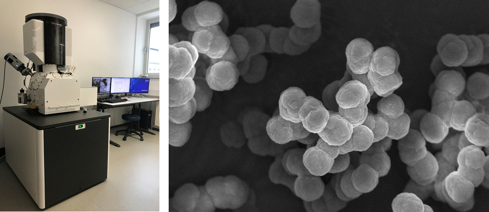
In the Center we have four scanning electron microscopes: JEOL JSM-5800 (located at IJS, Podgorica), JEOL JSM-7600F, Thermo Fisher Verios 4G HP and Thermo Fisher Quanta 650.
From left to right: JSM-5800, SE image LaB6, SE image W source, eye of a fly. JEOL JSM-5800 *IJS Podgorica
SE, BE detectors Oxford Instruments ISIS 300 EDS digital imaging From left to right: JSM-7600F, SE Image ZnO (author Mateja Podlogar, IJS), SE Image MoS2, BSE image of cheramics and dust. Jeol JSM-7600F
In-Lens Thermal FEG SEI, LEI detectors BE detectors (RIBE, RBEI) INCA Oxford 350 EDS SDD (20mm2) INCA Wave 500 spectrometer XENOS XeDraw 2 e-lithograph EBSD, Channel 5, Oxford Instruments r-filter for contaminating SE in BE signals prechamber IR CCD camera Thermo Fisher Verios 4G HP
Schottky FEG with a monochromator piezo stage movement automatic prechamber Nav-Cam inside the chamber IR CCD camera ETD in the chamber TLD in the column MD in the column for BSE ICD in the column for BSE DBS in the chamber for BSE STEM 3+ (BF, DF, HAADF) EDS, Oxford Instruments, AZtec Live, Ultim Max SDD 65 mm2 From left to right: Quanta 650, SE mirror image , SE image of particles, fibers and cheramics. Thermo Fisher Quanta 650 ESEM
differential pumping for three vacuum modes (HiVac, LoVac and ESEM mode) W thermionic source huge chamber, for 16 samples and 65nm movement in the Z axis Nav-Cam IR CCD camera ETD in the chamber CBSD on the objective lens LFD for LoVac GSED for ESEM mode two PLA apertures (EDS v LoVac) EDS, Oxford Instruments, AZtec Live, Ultim Max SDD 40 mm2


