

... Digital fields, Industry and Space, as well as in Food, Bioeconomy, Natural Resourses, Agriculture and the Environment.
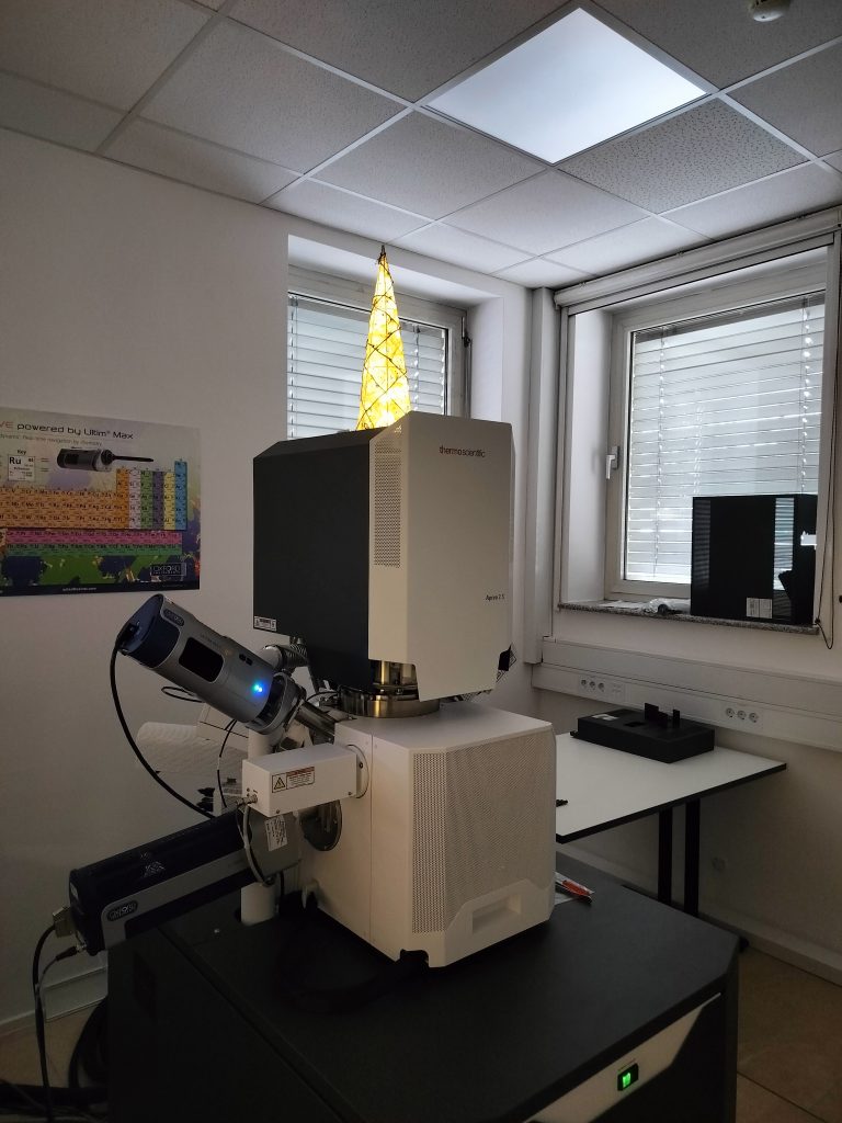
Advanced electron microscopy techniques enable the comprehensive characterization of inorganic materials in terms of their structural, morphological, and chemical properties. While scanning electron microscopy (SEM) enables such investigations at the micron level, transmission electron microscopy (TEM) enables the investigation of materials at the subatomic level and enables analyses such as grain boundary investigations, inclusions, the determination of planar defects, and dislocations in various minerals, and during in-situ experiments. The use of electron microscopy is also expanding into environmental investigations as an analytical technique for the structural, morphological, and chemical characterisation of natural materials in the environment, and potential inorganic and organic contaminants in both soil and water, and for the analysis of sensor materials for environmental applications.
Summer reading about science and a little around it.
Read the latest issue of IJS News.
https://ijs.si/ijsw/Novice%20IJS (pdf. file)

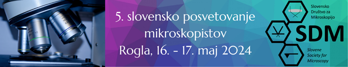
The main purpose of the conference is to encourage collaboration between younger and well-established Slovenian researchers and other users of microscopic techniques working in the fields of life science, material science, and industry. In addition to presenting the latest achievements in microscopy, the conference is also intended to showcase the activities of various groups using microscopic techniques at all levels of resolution.
The registration fee is 100 EUR for members of the Slovenian Society for Microscopy and 150 EUR for non-members, including participation in the symposium, abstract book, coffee breaks, and lunch on Friday. We have negotiated a special rate for accommodation with Hotel Superior on Rogla, including full board. When making a hotel reservation (reservation email: rogla@unitur.eu), please mention that you will be attending the SDM 2024 meeting.
Registrations, submission of contributions, and more information about the conference can be found on the website: https://www.mikroskopsko-drustvo.si/posvet/.
The deadline for abstract submissions is April 1, 2024, and the hotel reservation deadline is May 2, 2024.
See you at Rogla!