

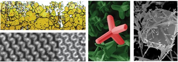
To end the summer, we want to share with you our collection of marine samples. Analysis was done on a Thermo Fisher Verios and Quanta 650 microscope.


CEMM is announcing a new professional/research position in the field of scanning electron microscopy and a new professional/research position in the field of transmission electron microscopy.
You are invited to submit applications.
Center for Electron Microscopy and Microanalysis, CEMM at Jozef Stefan Institute, IJS, calls for new candidates for expert/research works. The candidate will mainly work with the equipment for electron microscopy and sample preparation.
Employment will be made for a three months trial period full time with the possibility for extension to 12 months and afterwards to permanent employment. The candidates with already acquired knowledge in electron microscopy will be considered preferably.
Candidates can get additional information on (01)4773341 and/or 041743265, or via email: miran.ceh@ijs.si.
Written applications should be sent, together with the CV and copies of documents proving the expertise and education, to miran.ceh@ijs.si until October 31, 2023.
In May 2023 the third Advanced School on Scanning Transmission Electron Microscopy, AdSTEM3 was organized in Piran. The workshop was targeted at doctoral students, post-doctoral researchers and also already experienced microscopists and covered various topics on 4D STEM imaging. The conference included invited lectures by renowned professors. Emphasis was on the implementation and applications of various techniques resulting from sensitive direct electron detectors. 4D STEM, data-evaluation approaches and the choice of the appropriate evaluation software were presented. More information about the conference is available at the link.

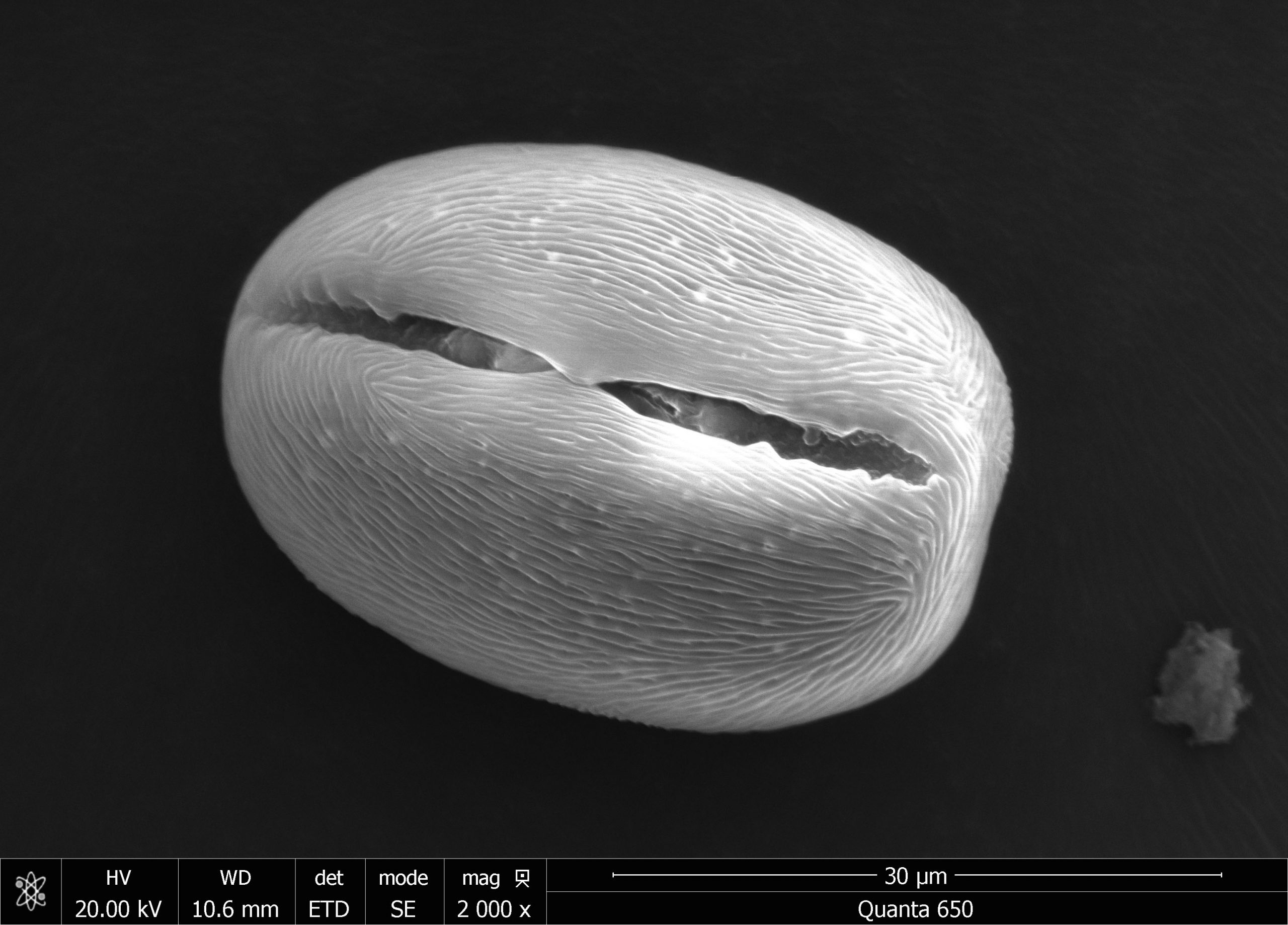
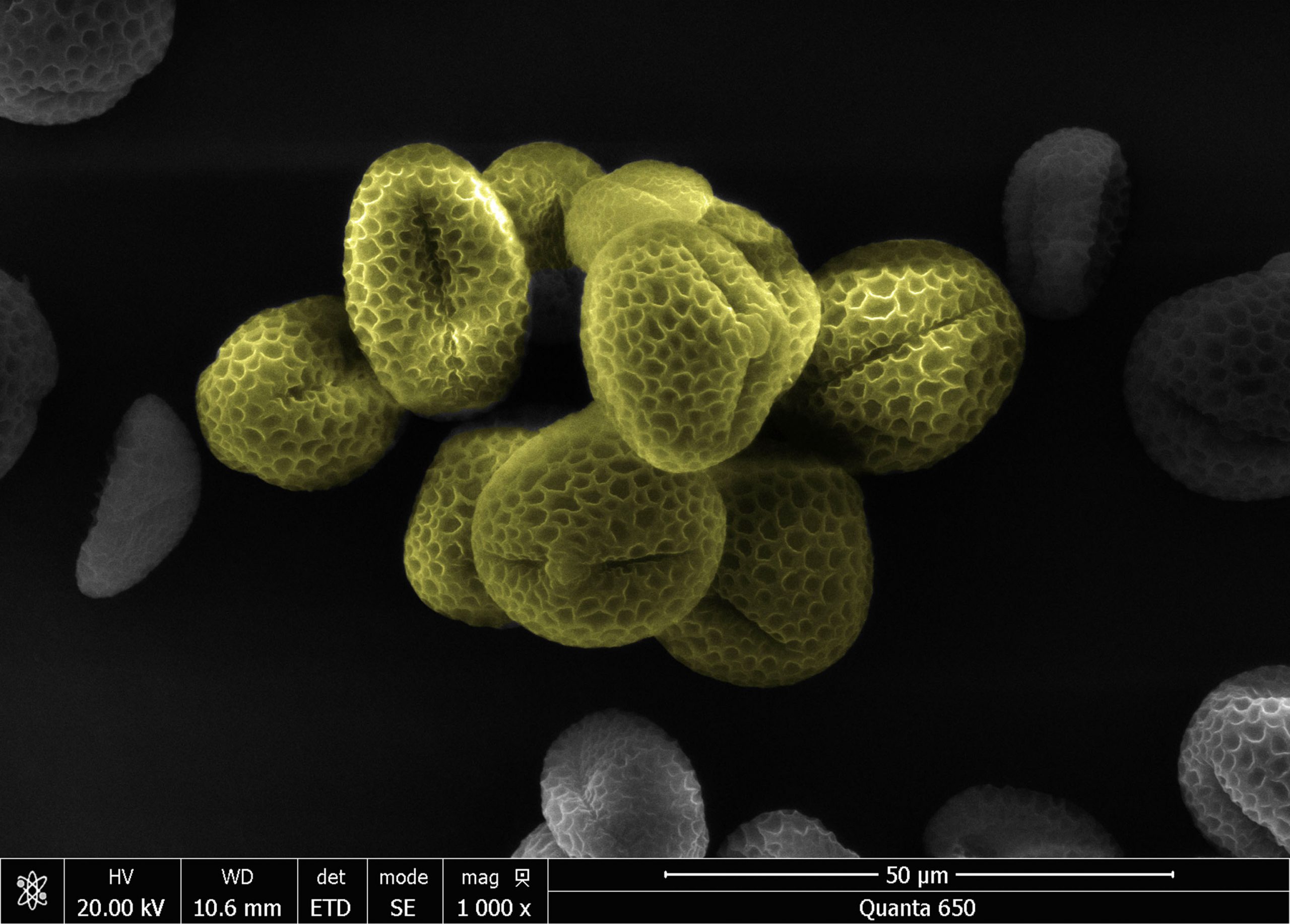
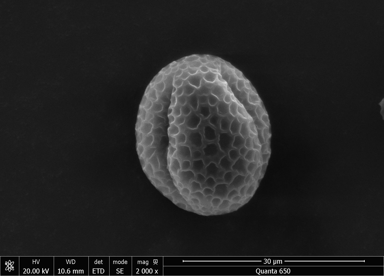
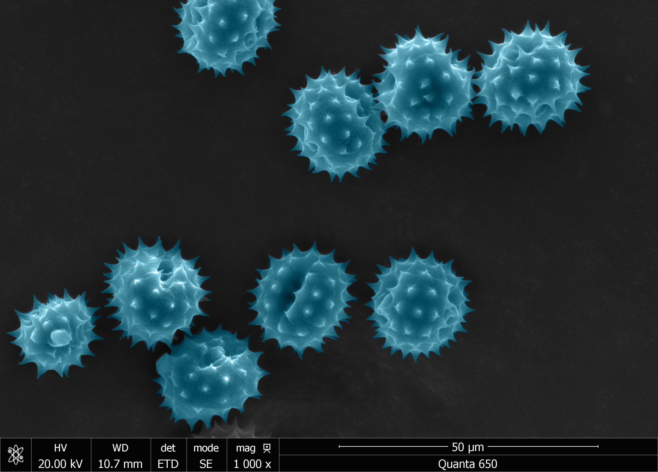
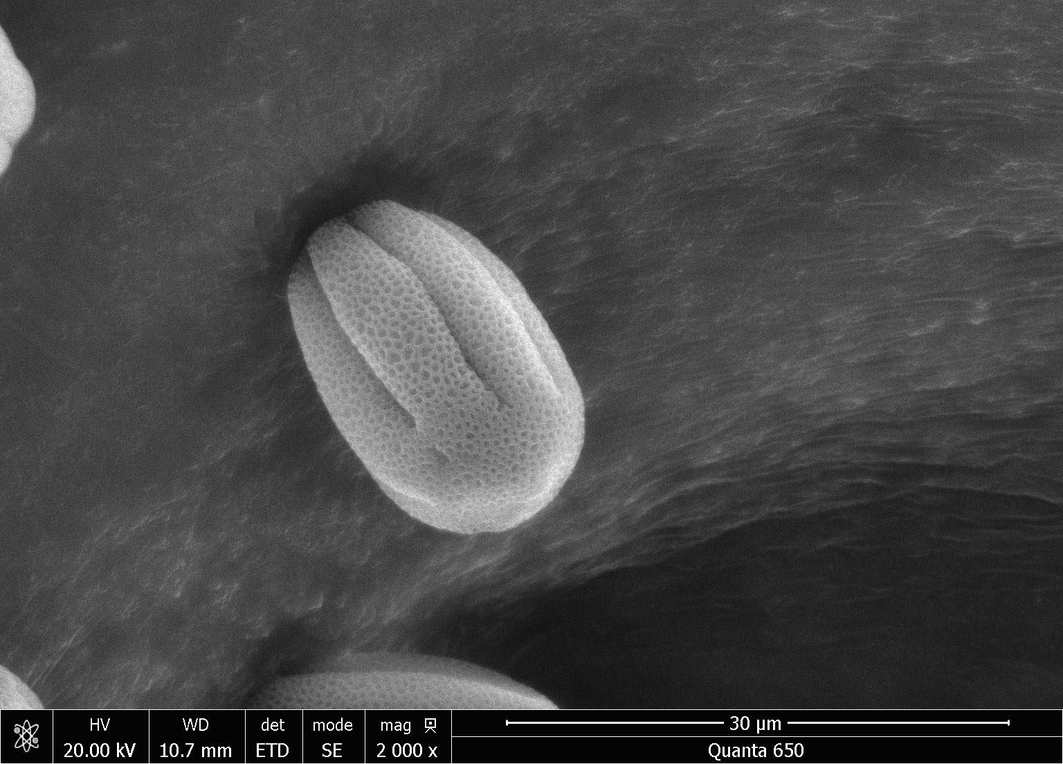
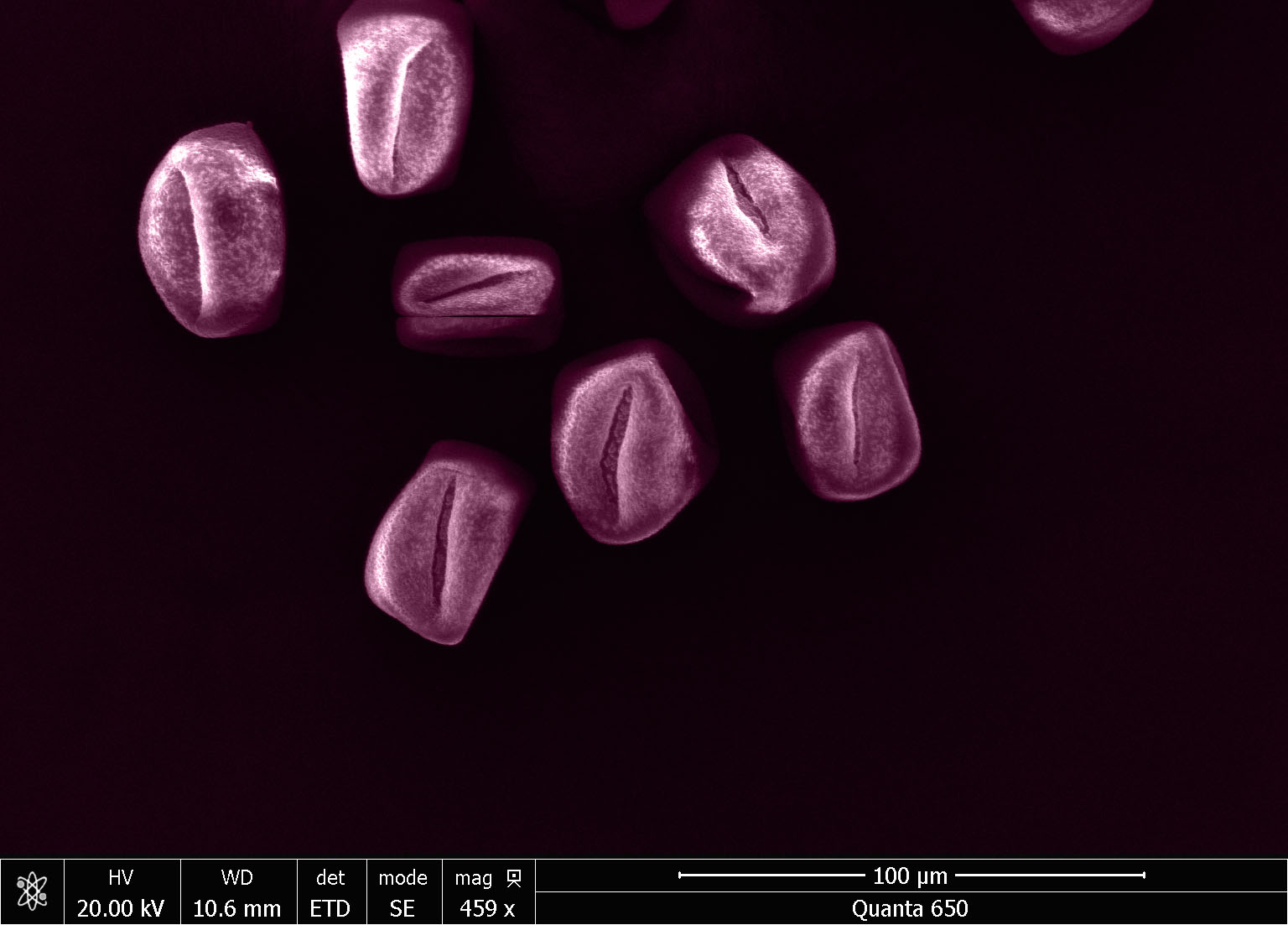
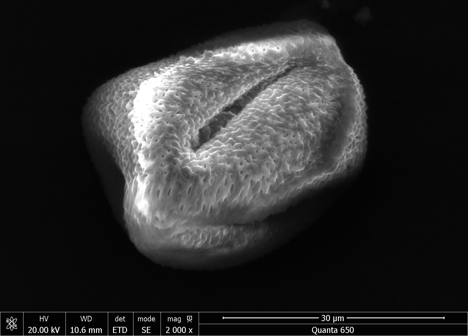
In the spirit of spring, we took some spring-harbinger plants under the microscope. Photographs of pollen were taken on Thermo Fisher Quanta 650 ESEM microscope. Images taken at lower magnifications are post-processed/colored with Adobe Photoshop.


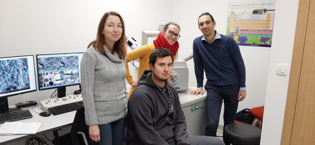
In December we hosted an expert on SEM Quanta 650 from Thermo Fisher at CEMM.
The following microscope operations were covered:
And advanced methods:


